Abdominal Anatomy : The Aorta - Branches - Aortic Arch - TeachMeAnatomy. 5 name the nine abdominal regions and their main contents. Divided into 9 regions by two vertical and two horizontal imaginary planes. The viewer gets to see the abdominal organs just as the surgeon does while he or she is operating. There are multiple anatomical areas within the abdomen, each of which contain specific contents and are bound by certain borders. Abdomen, in human anatomy, the body cavity lying between the chest or thorax above and the pelvis below and from the spine in the back to the wall the abdominal organs are supported and protected by the bones of the pelvis and ribcage and are covered by the greater omentum, a fold of peritoneum.
5 name the nine abdominal regions and their main contents. Transversus abdominis muscle internal abdominal oblique muscle rectus abdominis muscle external abdominal oblique muscle pyramidalis muscle. Level of l5, near transtubercular plane anatomy ileum, rectus abdominis muscle, ileocecal junction, cecum, internal abdominal oblique muscle, external abdominal oblique muscle, psoas major muscle, iliacus muscle, body of l5 vertebra. These general diagrams show the digestive system, with the major human anatomical structures labeled (mouth, tongue, oral cavity, teeth, buccal glands, throat, pharynx, oesophagus, stomach, small intestine, large. The abdominal cavity is an ovoid space bounded cephalad by the diaphragm and inferior thoracic margin, caudally by the pelvic brim, posteriorly by the lumbar spine along with quadratus lumborum, psoas major and iliacus, and.
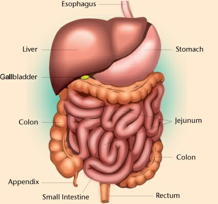
Abdominal Cavity - Definition and Organs | Biology Dictionary from biologydictionary.net In order to find the right training and to perform the exercises properly, it is important to know what are the abdominal muscles. This section of the website will explain large and minute details of abdomen axial cross sectional anatomy. The abdomen (colloquially called the stomach, belly, tummy or midriff) is the part of the body between the thorax (chest) and pelvis, in humans and in other vertebrates. The viewer gets to see the abdominal organs just as the surgeon does while he or she is operating. The xiphoid process and costal. But with the use of smart technology, you can learn faster and master abdomen anatomy in no time! Common incisions and closure techniques, and prevention and management of wound complications, are discussed elsewhere. The stomach, the small intestine (jejunum and ileum), the large intestine (colon), the liver, the spleen, the gallbladder, the pancreas, the uterus, the fallopian.
The abdomen refers to the region between the pelvis (pelvic brim) and the thorax (thoracic diaphragm) in vertebrates, including humans.
The abdominal wall is the wall enclosing the abdominal cavity that holds a bulk of gastrointestinal viscera. Two layers in abdomenfatty superficial layer (camper's fascia)deeper membranous layer (scarper's fascia). Respiratory muscle training online course: Connections between the left and the middle colic the gonadal arteries cross the abdominal ureters approximately halfway between the pelvic inlet and the renal pelvis. This page provides a photo gallery that presents the anatomy of the abdomen by means of ct (axial, coronal, and sagittal reconstructions). Windham was previously a surgical oncologist in the sarcoma program of the h. The stomach, the small intestine (jejunum and ileum), the large intestine (colon), the liver, the spleen, the gallbladder, the pancreas, the uterus, the fallopian. Abdominal anatomy seen on ct. Simple, easy notes for quick revision of important questions. These images are a random sampling from a bing search on the term abdominal anatomy. click on the image (or right click) to open the source website in a new browser window. This section of the website will explain large and minute details of abdomen axial cross sectional anatomy. This muscle forms the anterior and lateral abdominal wall. Radiology basics of abdominal ct anatomy with annotated coronal images and scrollable axial images to help medical students and junior doctors learning anatomy.
Transversus abdominis muscle internal abdominal oblique muscle rectus abdominis muscle external abdominal oblique muscle pyramidalis muscle. Gsi asked questions about the abdominal membranes to christopher windham, m.d. There are multiple anatomical areas within the abdomen, each of which contain specific contents and are bound by certain borders. A good amount of area is covered by the abdominal wall. But with the use of smart technology, you can learn faster and master abdomen anatomy in no time!
Anatomy of the Male Abdomen/Pelvis | Trial Exhibits, Inc. from cdn.trialexhibitsinc.com This is a laparoscopic tour of abdominal cavity anatomy. Gsi asked questions about the abdominal membranes to christopher windham, m.d. Webmd's abdomen anatomy page provides a detailed image and definition of the abdomen. The abdomen is comprised primarily of the digestive tract and other accessory organs which assist in digestion, the urinary system, spleen, and the abdominal. Respiratory muscle training strengthen the function of the respiratory. Transversus abdominis muscle internal abdominal oblique muscle rectus abdominis muscle external abdominal oblique muscle pyramidalis muscle. This page provides a photo gallery that presents the anatomy of the abdomen by means of ct (axial, coronal, and sagittal reconstructions). A good amount of area is covered by the abdominal wall.
The above lines intersect and divide the abdomen into nine regions (clockwise from the top)
A good amount of area is covered by the abdominal wall. This muscle forms the anterior and lateral abdominal wall. Two layers in abdomenfatty superficial layer (camper's fascia)deeper membranous layer (scarper's fascia). The vascular anatomy distal to the middle colic artery and near the splenic flexure is variable. Level of l5, near transtubercular plane anatomy ileum, rectus abdominis muscle, ileocecal junction, cecum, internal abdominal oblique muscle, external abdominal oblique muscle, psoas major muscle, iliacus muscle, body of l5 vertebra. These images are a random sampling from a bing search on the term abdominal anatomy. click on the image (or right click) to open the source website in a new browser window. Windham was previously a surgical oncologist in the sarcoma program of the h. The xiphoid process and costal. The abdomen contains many vital organs: There are multiple anatomical areas within the abdomen, each of which contain specific contents and are bound by certain borders. The above lines intersect and divide the abdomen into nine regions (clockwise from the top) Abdomen, in human anatomy, the body cavity lying between the chest or thorax above and the pelvis below and from the spine in the back to the wall the abdominal organs are supported and protected by the bones of the pelvis and ribcage and are covered by the greater omentum, a fold of peritoneum. Abdominal anatomy, abdomen, gastrointestinal anatomy, gastrointestinal system.
This muscle forms the anterior and lateral abdominal wall. Windham was previously a surgical oncologist in the sarcoma program of the h. The vascular anatomy distal to the middle colic artery and near the splenic flexure is variable. Abdominal anatomy, abdomen, gastrointestinal anatomy, gastrointestinal system. 6 write the origin, insertion and nerve supply of muscles of anterior abdominal wall.
Anterior abdominal muscles 1: rectus abdominis | Acland's ... from aclandanatomy.com Connections between the left and the middle colic the gonadal arteries cross the abdominal ureters approximately halfway between the pelvic inlet and the renal pelvis. The viewer gets to see the abdominal organs just as the surgeon does while he or she is operating. Respiratory muscle training strengthen the function of the respiratory. Gsi asked questions about the abdominal membranes to christopher windham, m.d. Respiratory muscle training online course: The abdomen contains many vital organs: The abdomen is comprised primarily of the digestive tract and other accessory organs which assist in digestion, the urinary system, spleen, and the abdominal. Abdomen, in human anatomy, the body cavity lying between the chest or thorax above and the pelvis below and from the spine in the back to the wall the abdominal organs are supported and protected by the bones of the pelvis and ribcage and are covered by the greater omentum, a fold of peritoneum.
The abdomen refers to the region between the pelvis (pelvic brim) and the thorax (thoracic diaphragm) in vertebrates, including humans.
Abdominal surface anatomy can be described when viewed from in front of the abdomen in 2 ways: The abdomen contains many vital organs: This page provides a photo gallery that presents the anatomy of the abdomen by means of ct (axial, coronal, and sagittal reconstructions). The abdomen refers to the region between the pelvis (pelvic brim) and the thorax (thoracic diaphragm) in vertebrates, including humans. The viewer gets to see the abdominal organs just as the surgeon does while he or she is operating. The stomach, the small intestine (jejunum and ileum), the large intestine (colon), the liver, the spleen, the gallbladder, the pancreas, the uterus, the fallopian. Divided into 9 regions by two vertical and two horizontal imaginary planes. Anatomy of the abdominal wall. 5 name the nine abdominal regions and their main contents. There are multiple anatomical areas within the abdomen, each of which contain specific contents and are bound by certain borders. Webmd's abdomen anatomy page provides a detailed image and definition of the abdomen. The abdomen (colloquially called the stomach, belly, tummy or midriff) is the part of the body between the thorax (chest) and pelvis, in humans and in other vertebrates. Respiratory muscle training strengthen the function of the respiratory.
Share :
Post a Comment
for "Abdominal Anatomy : The Aorta - Branches - Aortic Arch - TeachMeAnatomy"


Post a Comment for "Abdominal Anatomy : The Aorta - Branches - Aortic Arch - TeachMeAnatomy"